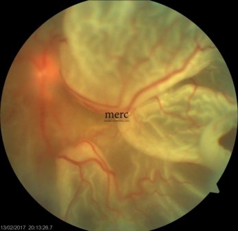
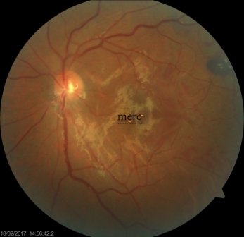
Retinal Detachment Definition:
Retinal detachment refers to the separation of the retina from the underlying tissues namely the RPE and choroid.
The retina is the light-sensitive inner lining of the eye and remains apposed (loosely attached) to its underlying layers namely the choroid. The deeper layers of the retina receive its blood supply from the choroid. A retina detachment is therefore serious as the retinal blood supply stops and so does the oxygen supply. If a retina detachment is not corrected quickly, it can produce permanent damage. Fortunately, a retinal detachment has clear warning signs. Early diagnosis and treatment can save your vision. if you suspect you may have the retinal detachment, contact a retina specialist today.
Retinal Detachment Symptoms:
It is a painless eye condition. Following signs should alert you to visit a retinal specialist to rule out a retinal detachment right now
- a sudden appearance of many floaters - tiny particles like spots/hair/strings that float in your vision and move with eye movements
- sudden flashes of light - a bright spark like lightning which can be perceived even with eyes closed
- shadow or curtain over part of the vision which progressively worsens
How does a retina detachment occur?
The common retinal detachment occurs when the gel of the eye (vitreous) leaks through holes/tears in the retina thereby lifting it from the choroid
Why does the retina tear?
As one ages, the gel that fills the eye shrinks/collapses and becomes more liquid. Eventually the vitreous may separate from the retina a process called posterior vitreous detachment (PVD). When the vitreous peels of from the retina, it may sometimes pull it forcefully resulting in a retinal tear
Detached Retina Risk Factors:
- Myopia- High minus numbers
- A family history of retinal detachment
- History of RD in one eye
- Any eye surgery including cataract and LASIK
- Laser Capsulotomy (YAG)
Retinal Detachment Surgery:
The goal is to get the retina back quick in place so that it starts getting its blood supply and oxygen. A retinal detachment is fixed by a retina specialist by simple & advanced surgical techniques
Scleral Buckle
- In this technique, the retinal tear is fixed by an "EXTERNAL" approach ( without entering the eye). In scleral buckling, the tear is sealed using a freezing technique called “CRYOTHERAPY’ and then supported by a piece of SOLID SILICONE BUCKLE. It is a technique that requires great skill and clinical acumen
Vitreo Retinal Surgery
- In this technique the retinal tear is fixed by an "INTERNAL" approach It uses sophisticated machines and instruments (MIVS - MICROINCISION VITREOUS SURGERY) to remove the gel of the eye, the vitreous. The tear is then sealed with the LASER. The retina is then supported with either SILICONE OIL or INTRAOCULAR GAS injection. The patient is required to maintain the face, down position for few days after the surgery


